


Changes in condylar position after orthognathic surgery and its correlation with temporomandibular symptoms (TMD)- a prospective study
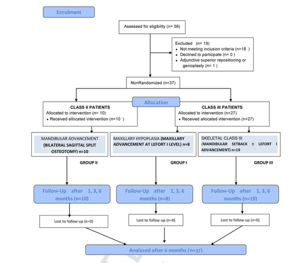
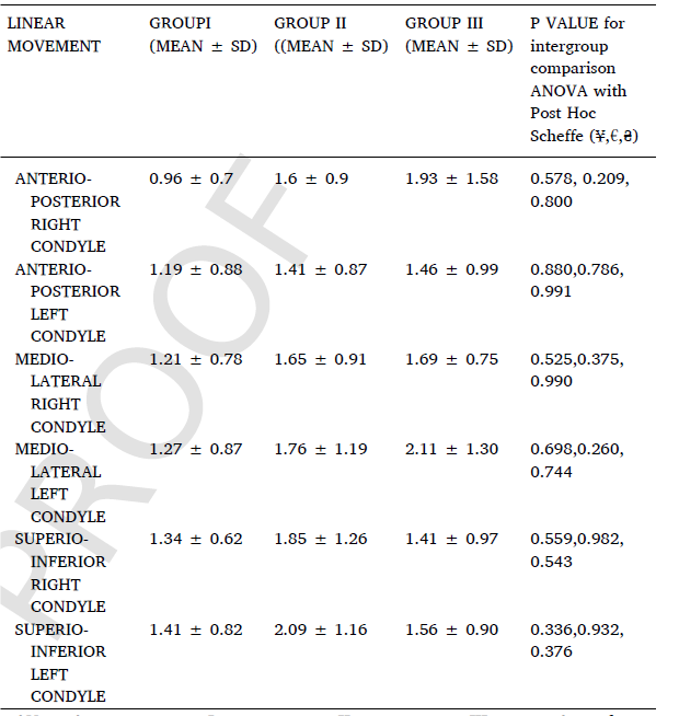
| Abstract | The aim of the study was to assess the changes in the condylar position after orthognathic surgery (OGS) and its effect on temporomandibular disorders (TMD). A total of 37 dentofacial deformity patients included in the study who had undergone OGS were divided into three groups: Group I, Le Fort I maxillary advancement; Group II, bilateral sagittal split osteotomy (BSSO) mandibular advancement ± Le Fort I; and Group III, BSSO mandibular setback ± Le Fort I. Patients were evaluated clinically using Diagnostic Criteria for TMD and by radiography preoperatively and 6 months postoperatively. The positional changes in condyle were correlated with signs and symptoms of TMD. A total of 37 patients in three groups (Group I, 8 patients; Group II, 10 patients; and Group III, 19 patients) were evaluated. Overall, condyles had anterio-medio-inferior movement with 7 of 8 patients in Group I, 6 of 10 patients in Group II and 13 of 19 patients in Group III having ≤2 mm displacement. In angular changes, inward-anterio-medial movement was observed with 6 of 8 patients in Group I; about 5 of 10 patients, and 10 of 19 patients in Group II and III respectively had ≤5° change. Intragroup and intergroup comparisons showed insignificant changes in TMD and linear/angular movement (p ≥ 0.05). Pearson correlation coefficient was found to be nonsignificant on the radiographic and clinical comparison (p ≥ 0.05). Intrarater reliability (Kappa value) was found to be 0.83, confirming the results. Within the limitations of the study it seems that there are minimal linear and angular changes in condyle after orthognathic surgery that were not responsible for the development of temporomandibular disorders in the postoperative course. |
| Faculty |
Prof. Parveen Kalra
|
|
parveenkalra@pec.edu.in
|
|
| Collaborations | PGIMER, Chandigarh |
| More Information | https://doi.org/10.1016/j.jcms.2022.12.003 |